

Applications of Panoramic Endoscopic Imaging; Lens Manufacturer
1 Capsule Endoscope
Currently, numerous manufacturers worldwide can provide capsule endoscopes with large viewing angles. Capsule endoscopes equipped with a single ultra-wide-angle lens typically have a field of view (FOV) between 156° and 170°, resulting in similar visible ranges. Differences mainly lie in frame rate, communication method, battery life, and other aspects. In recent years, with advancements in miniaturization technology and reduced power consumption of related components, manufacturers have gradually begun integrating more cameras into capsule endoscopes.
Medtronic, based on its PillCam SB series, developed a capsule endoscope for Crohn's disease featuring two cameras, one on the front and one on the rear. Each camera has a 168° FOV. Similarly, IntroMedic's MC2000 series also incorporates two cameras (front and rear), each with a 170° FOV, providing simultaneous forward and backward views. However, the frame rate decreases from the original 6 frames per second (fps) for a single-camera system to 3 fps per camera, ensuring manageable data storage and transmission.
Differing from other capsule endoscopes, the device developed by CapsoVision incorporates four cameras distributed around its sidewall, as shown in Figure 16(a). By stitching the images from these four cameras, 360° panoramic imaging of the intestine can be achieved, providing an excellent view for observing folds, polyps, and other lesions on the intestinal wall. With ongoing improvements in communication modules, battery life, and other technologies, integrating more cameras and achieving better image quality will be the main direction for capsule endoscope development.
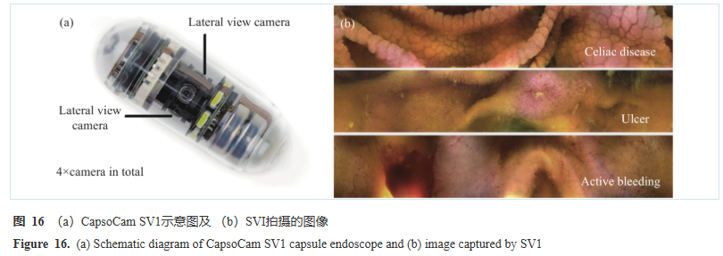
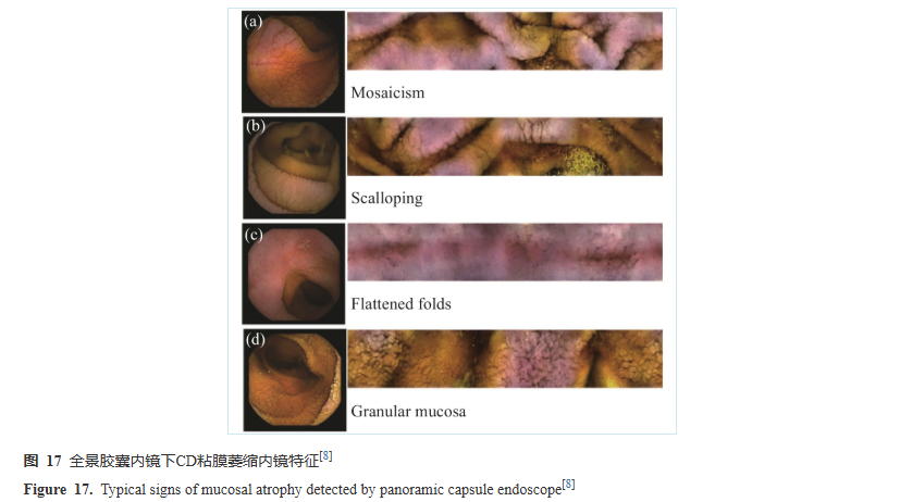
Capsule endoscopes are now extensively used and play an important role in diagnosing and treating conditions such as obscure gastrointestinal bleeding (OGIB), Crohn's disease, and complicated celiac disease. Figure 16(b) shows images captured by the CapsoCam SV1 for various common conditions, including celiac disease, ulcers, and active bleeding. In one comparative study between CapsoCam SV1 and PillCam SB3 involving 153 patients with OGIB, the results showed that the SV1, with its panoramic view, could observe richer pathological information and detect more cases of bleeding. Regarding physician satisfaction, 95% were satisfied with the capsule system and assessment software, while treatment-related adverse events/serious adverse events were 17.9%/1.3%. Overall, patient acceptance of the SV1 was high, indicating broad application prospects in outpatient settings. In the diagnosis and management of Crohn's disease, using the panoramic-view CapsoCam SV1, physicians observed numerous erosions and ulcers from the distal duodenum to the terminal ileum in patients suspected of having Crohn's disease, suggesting practical improvements in lesion classification and differential diagnosis. Based on these features, the patient was diagnosed with extensive small bowel Crohn's disease and improved with specific treatment. For celiac disease, using the SV1 effectively detected small bowel atrophy, showing good sensitivity and specificity compared to histology. Figure 17 shows an image of the small bowel area in a CD patient taken with the SV1, capable of detecting four endoscopic features of mucosal atrophy, providing significant guidance for diagnosing CD.
2 Colorectaloscope
Similar to capsule endoscopes, colorectaloscopes utilizing panoramic imaging are also gaining widespread application.
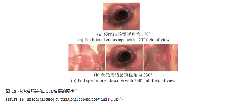
Colorectal cancer (CRC) is currently the third most commonly diagnosed cancer. According to related statistics, 60% of CRC cases originate from adenomas, and 35% originate from sessile serrated adenomas/polyps. Colonoscopy aims to detect and remove these precancerous polyps at an early stage. However, conventional colonoscopy still misses a significant number of polyps, with a pooled miss rate for polyps of any size being 22%. In comparative imaging of colonic polyps using traditional colonoscopy versus the FUSE system, as shown in Figure 18, polyps on the sidewalls missed during traditional colonoscopy can be observed in the lateral views provided by FUSE, thereby reducing the miss rate, decreasing physician working time, and improving efficiency.
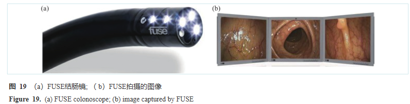
Recent related research has proposed behind folds visualizing (BFT) technologies and techniques aimed at improving the adenoma detection rate (ADR). The Full Spectrum Endoscopy (FUSE) system, developed by Endo-Choice, adds two lateral-viewing lenses to a single forward-viewing lens, expanding the FOV to 330°. Images captured during its operation are displayed on different screens. Olympus's EWAKE colonoscope adopts a similar scheme, comprising one standard 147° forward-viewing lens and two additional 42.5° lateral rear-viewing lenses. However, it synthesizes the views from all lenses and displays them as a single endoscopic image on the monitor. The Third Eye Panoramic device, developed by Avantis Medical, consists of two side-view cameras that can be attached to the end of a standard colonoscope, producing three images that can be projected onto the screen, extending the viewing angle to over 300°. Although some studies indicate that using these technologies does not significantly improve ADR compared to traditional colonoscopes, employing BFT can enhance the detection of non-advanced polyps and lesions, effectively reducing the risk of missing non-advanced adenomas. Furthermore, for less experienced colonoscopists, using BFT provides a richer field of view, and the benefits are more pronounced compared to when used by highly experienced physicians.
Previous:MOTION DV LENS