

Endoscopic Lenses: The Doctor's "Magic Eye"
Endoscopic Lenses: The Doctor's "Magic Eye"
From candlelit rigid scopes in the late 18th century to today’s 4K ultra-HD electronic endoscopes, this technology has undergone a revolutionary evolution, becoming the cornerstone of minimally invasive medicine. This article will guide you through the fascinating world of endoscopic lenses—from historical developments to modern applications, from rigid scopes to flexible ones, and finally to AI-assisted future trends—revealing how this technology enables doctors to diagnose and treat diseases with precision without opening the human body.
1. Evolution of Endoscopic Lenses: From Candlelight to Electronic Signals
The history of endoscopy dates back to the late 18th century, when German physician Philip Bozzini (1804) invented a primitive cystoscope equipped with candlelight illumination, attempting to observe internal human structures. However, limited by the light source technology and materials science of the time, these early rigid endoscopes had numerous issues: narrow field of view, insufficient illumination, tissue damage risks, and even burns. It wasn’t until 1879 that German doctor Nitze replaced candlelight with Edison’s electric bulb, solving some illumination challenges.
In 1930, German physician Lamm discovered that light could still be transmitted through bundled micrometer-diameter fiber strands even when bent—a breakthrough that laid the foundation for fiber-optic endoscopes. In 1957, Hirschowitz and his team demonstrated the first fiberoptic endoscope for examining the stomach and duodenum, marking the birth of flexible endoscopes. The greatest advantage of fiberoptic endoscopes lies in their softness and flexibility, which greatly reduces patient discomfort while enabling early detection of tiny lesions like cancer and ulcers. However, the fragility of optical fibers and image transmission issues like black spots limited their lifespan.
The real leap in endoscopic technology occurred in 1983 when Welch Allyn (USA) and Japanese companies developed electronic endoscopes—the third generation of endoscopes. These replaced optical fibers with CCD sensors, converting optical images into TV signals displayed on screens. This revolution made image storage, reproduction, remote consultation, and computer management possible. Image clarity and resolution improved dramatically—from the initial 10,000 pixels (fiberscopes) to 40,000–100,000 pixels (early electronic scopes) and now up to 8 million pixels (4K lenses). This is akin to jumping from blurry black-and-white photos to 4K ultra-HD TVs, allowing doctors to see unprecedented details inside the human body.
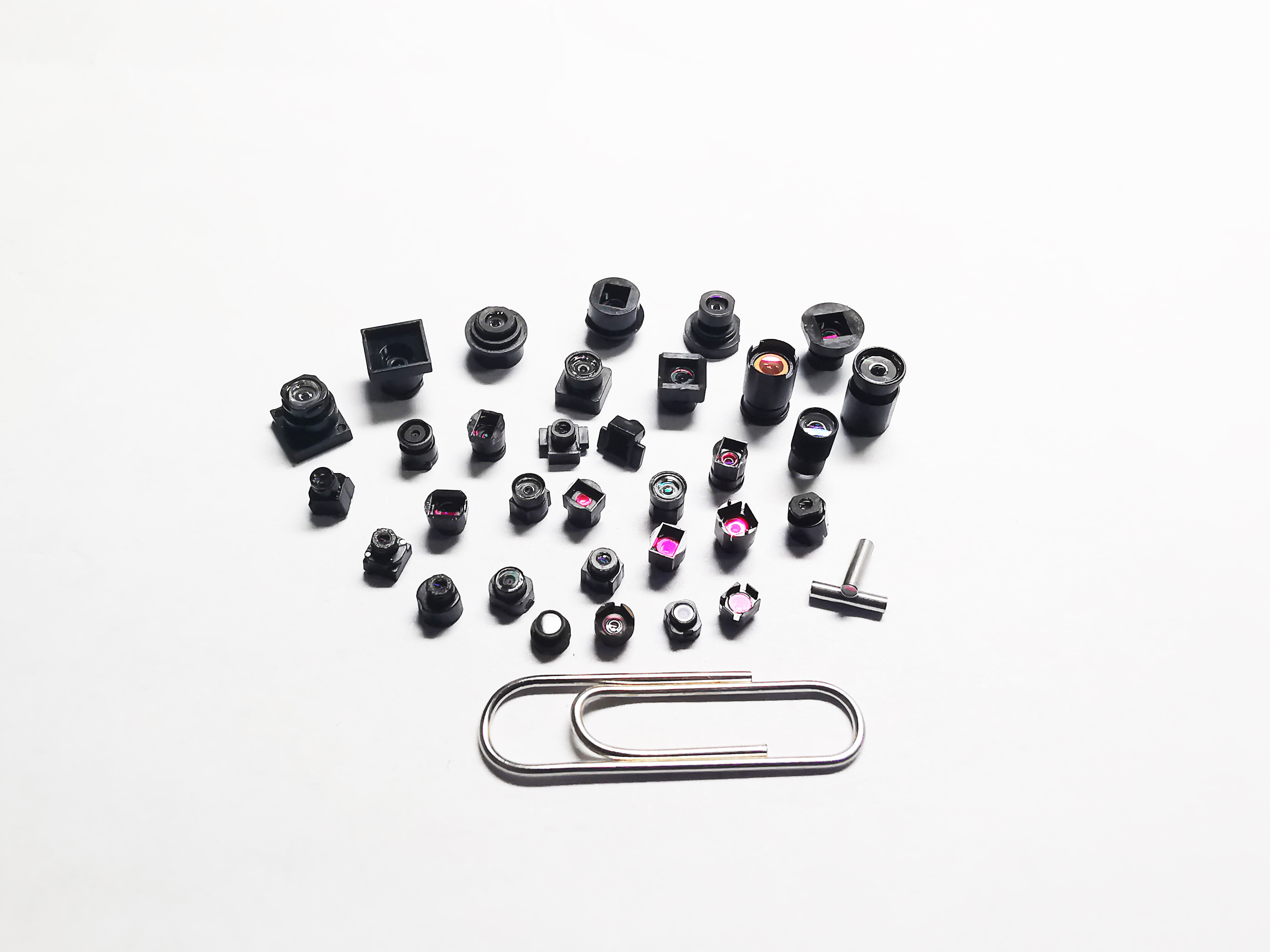
2. Types and Key Parameters of Endoscopic Lenses: How to Choose the Right Lens
Endoscopic lenses vary by type and application scenario. They are mainly divided into four categories: rigid endoscopic lenses, flexible endoscopic lenses, fiberoptic lenses, and electronic lenses, each with unique advantages and use cases.
Rigid endoscopic lenses typically consist of multiple optical lens groups that transmit images via optical refraction and reflection principles. Their diameter ranges from 5–12mm, with fixed field angles (e.g., 30°, 70°), short depth of field, and high resolution. Rigid scopes excel in sharp imaging and can be equipped with multiple working channels, ideal for precise minimally invasive surgeries. For example, laparoscopic surgeries often use 30° field-angle lenses because they clearly present organ layer structures, helping doctors judge tissue spacing.
Flexible endoscopic lenses use optical fibers or electronic sensors, with the key feature being the operator-controllable bending tip that expands applications. Their diameter is finer (e.g., ~12.6mm for gastroscopes), with large bending angles (dual-axis control), long depth of field, and flexible field angles (e.g., 0°, 30°, 70°). Flexible scopes resemble nimble snake-like robots, freely navigating complex internal cavities—perfect for deep observation in the digestive and respiratory tracts. For instance, colonoscopies require long focal lengths and large depth of field to maintain clarity over long distances, while bronchoscopies demand 30° or 70° lenses to visualize bronchial branches.
Fiberoptic lenses transmit images via optical fibers, offering wide field angles (10,000 pixels) and susceptibility to black spots, with shorter lifespans. Electronic lenses, however, use CCD or CMOS sensors to digitize images, achieving resolutions up to 1920×1080 or higher, with superior image quality. As technology advanced, CMOS sensors gradually replaced CCDs due to their lower power consumption, stronger anti-interference circuitry, and high integration, becoming the mainstream choice.
When selecting lenses, doctors consider multiple parameters:
|
Parameter |
Rigid |
Flexible |
Fiberoptic |
Electronic |
|
Diameter |
5–12mm |
2.8–12.6mm |
<6mm |
2.8–12.6mm |
|
Field Angle |
Fixed (e.g., 30°, 70°) |
Variable (0°, 30°, 70°) |
~140° wide-angle |
Variable (0°, 30°, 70°) |
|
Resolution |
High (up to 8MP) |
Medium (10K–100K pixels) |
Low (~10K pixels) |
High (1920×1080–3840×2160) |
|
Bending Angle |
Fixed |
Large (e.g., 180°) |
Medium |
Large (e.g., 180°) |
|
Depth of Field |
Short |
Long |
Long |
Adjustable |
|
Durability |
High |
Medium |
Low |
High |
3. Lens Materials and Manufacturing Innovations: Enhancing Medical Imaging Quality
Lens materials and manufacturing processes critically impact image quality. From early ordinary glass to modern sapphire and special alloys, materials science has significantly improved lens durability and optical performance.
Sapphire lenses, a recent innovation, are composed of aluminum oxide, second only to diamonds in hardness, with excellent wear and corrosion resistance. Sapphire lenses are as hard as diamonds but more transparent than regular glass, resisting scratches and impacts for long-term use. For example, SINGLON Medical’s 0.35mm ultra-thin endoscopic lens uses sapphire material, enabling access to microscopic ducts like tear glands and root canals—a domestic innovation.
Glass metallization is another breakthrough. Using laser-induced plasma-assisted ablation (LIPAA), researchers coat glass surfaces with metallic films, enhancing oxidation and corrosion resistance. This metallic layer acts as an "invisible armor," protecting lenses from disinfectants and bodily fluids to extend lifespan. For instance, DING Hongrun’s sapphire lenses, after metallization, improved oxidation resistance and surface hardness for harsher conditions.
Coating advancements also boosted optical performance. Sapphire glass with colorless anti-reflective coatings increased transmittance from 86.5% to 96.7%, acting as an "optical amplifier" to deliver clearer, truer images to doctors. Double-sided coatings offer 6% higher transmittance than single-sided ones, with better thermal stability, UV aging resistance, and wear resistance—ensuring stable performance in extreme conditions.
Manufacturing innovations have also driven miniaturization. Japanese companies developed ultra-fine gradient-index (GI) lenses as small as 0.1mm in diameter, reducing endoscope shaft sizes to under 1mm—half current mainstream products. This breakthrough enables endoscopes to access narrow anatomical regions like tear ducts, mammary ducts, and root canals, opening new diagnostic and therapeutic possibilities.
4. AI Assistance and Super-Miniaturization: Future Trends in Endoscopic Lenses
Endoscopic lens technology is undergoing a dual revolution with AI assistance and super-miniaturization, expanding applications and improving diagnostic and therapeutic precision.
AI-assisted endoscopy systems analyze image data in real time to identify potential lesions. For example, Morning Medical’s AI algorithms optimize image noise, enhancing clarity in low-light environments. Olympus Medical’s intelligent navigation system supports preoperative 3D modeling and intraoperative automatic vessel avoidance, upgrading surgical planning from "experience-driven" to "data-driven". AI acts as an experienced "imaging assistant," silently analyzing images and marking suspicious areas to reduce missed diagnoses while surgeons focus on operations.
Super-miniaturization is another key trend. SINGLON Medical’s 0.35mm ultra-thin lens is already used in dental root canal treatments, with future potential for cerebral vessels and nerve terminals. These ultra-fine lenses act as "medical spies," infiltrating the body’s narrowest cavities to capture cell-level HD photos, offering unprecedented microscopic views. For instance, its 0.35mm lens achieves a 0.5–120mm depth of field, broader than traditional lenses, capturing both micro and macro details simultaneously.
Disposable endoscopes are another growing direction. With CMOS chip localization and mature supply chains, single-use endoscope costs have dropped to around $1,000, promoting adoption in grassroots hospitals. Disposable lenses eliminate cross-infection risks and simplify cleaning processes, akin to "use-and-discard smartphones"—safe and convenient. In China, approved single-use endoscope registrations surged from 69 in 2022 to 366 in 2025, with urology products exceeding 50%—highlighting this trend’s momentum.
Fluorescence navigation is another highlight. Injecting contrast agents like indocyanine green (ICG) makes tumors and lymph tissues glow, enabling fluorescence endoscopes to precisely mark liver cancer margins for millimeter-level resection. Fluorescence endoscopes act as "night vision goggles," illuminating tumor boundaries in the dark to guide precise removal. Hisun Medical, manufacturing 70% of Stryker’s global fluorescence laparoscopes, achieves millimeter-level liver cancer margin marking.
5. Clinical Applications of Endoscopic Lenses: Comprehensive Support from Diagnosis to Treatment
Endoscopic lenses are not just for diagnosis but also widely used in minimally invasive treatments. From simple observation to complex surgeries, endoscopic lenses have become multifunctional "toolkits" in doctors’ hands.
In gastrointestinal disease checks, endoscopic lenses directly observe lesions like ulcers, inflammation, polyps, and tumors in the esophagus, stomach, duodenum, small intestine, and colon. For example, gastroscopy uses CCD sensors at the endoscope tip to capture cavity optical signals, allowing doctors to view gastric mucosa details and detect early cancers. Gastroscopy lenses act as "micro-detectives," uncovering invisible lesions to provide timely treatment advice.
In respiratory disease checks, bronchoscopes and laryngoscopes delve into lungs and throats, observing bronchial and vocal cord lesions. These lenses are like "respiratory explorers," guiding doctors through the body’s mysterious internal world. For instance, 30° or 70° bronchoscopes visualize bronchial branches to uncover hidden lesions.
In urological checks, cystoscopes and ureteroscopes directly inspect urinary system structures. Urological endoscopes act as "pipeline engineers," inspecting tubular organs like ureters and bladders for lesions. Fluorescence endoscopes in urology help identify tumor margins, improving surgical precision.
In laparoscopic surgeries, endoscopic lenses serve as both observation tools and surgical platforms. Doctors perform biopsies, hemostasis, and laser treatments via laparoscopes, integrating diagnosis and treatment. Laparoscopic lenses are "surgical commanders," providing visual information and operational channels to complete complex minimally invasive surgeries.
6. Conclusion: The Future of Endoscopic Lenses
From candlelit rigid scopes in the late 18th century to today’s AI-assisted 4K ultra-HD lenses, endoscopic technology has evolved revolutionarily from "seeing" to "penetrating". In the future, with AI, new materials, and optics deeply integrated, this "microscopic eye" will continue breaking human cognitive boundaries, benefiting more patients through precise, safe minimally invasive diagnostics and treatments.
AI assistance will transform endoscopic lenses from "passive observers" to "active assistants", enabling real-time lesion recognition, treatment suggestions, and even surgical decision-making. Super-miniaturization will explore the "last centimeter" of the human body, allowing endoscopes to enter narrower, more complex cavities for minimally invasive solutions. Disposable technology will drive inclusive healthcare, popularizing single-use endoscopes in grassroots hospitals and improving medical resource accessibility.
Endoscopic lenses are not just medical technological products—they are tools for exploring human mysteries. Their development reflects humanity’s relentless pursuit of health and showcases the immense potential of technology and medicine integration. With continuous technological advancements, endoscopic lenses will keep expanding our horizons, helping doctors treat diseases more accurately and safely, delivering better medical experiences to patients.
Next time you undergo an endoscopic examination, imagine how this magical lens becomes the doctor’s "magic eye," guiding them to explore your body’s secrets and safeguard your health. Though small, endoscopic lenses carry the future of medicine and the hope of life.